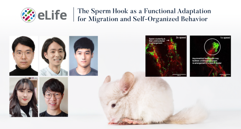
A research team affiliated with UNIST has made a groundbreaking discovery regarding the movement of mouse sperm, demonstrating how the hook-like structure at the sperm head aids in navigating the inner walls of the uterus.
Led by Professor Jung-Hoon Park from the Department of Biomedical Engineering at UNIST, this collaborative study also involved Professor Jae-Ick Kim from the Department of Life Sciences and Dr. Heungjin Ryu from Kyoto University. Utilizing advanced real-time deep tissue imaging enabled by a custom-built two-photon microscope, the team investigated sperm movement within the reproductive tract of the house mouse (Mus musculus, C57BL/6), providing unprecedented insights into their migration dynamics.
The primary aim of the study was to address two conflicting hypotheses regarding the function of sperm hooks in rodents. One hypothesis, referred to as “suspension cooperation,” suggested that sperm could enhance their speed in reaching eggs by linking their hook-like heads together, akin to a train. However, this phenomenon was not observed in the study’s findings.
Instead, the researchers revealed that mouse sperm use their hooks to interact with the epithelial lining of the reproductive tract, facilitating superior navigation through the uterus and oviduct. The sperm hook serves a dual purpose: it not only assists in anchoring the sperm to the epithelium but also enhances interaction with the reproductive tract, ultimately improving the sperm’s ability to overcome the challenges posed by strong fluid flows.
For the first time, the study observed organized, synchronized swimming movements among sperm, with heads aligning in a singular direction while their tails moved in unison, similar to synchronized swimmers. The research team proposed that the anchoring capability of the sperm hook is a key factor in achieving this coordination, providing the sperm with an evolutionary advantage for efficient migration.
“The train hypothesis, which has been observed only in two-dimensional culture dishes, could not be substantiated in our three-dimensional in vivo observations,” explained the research team. “While we did see small clusters moving together in a train-like formation, their speed was not comparable to that of individual sperm.” They emphasized the necessity for further research to gather statistically significant data to fully evaluate the train hypothesis.
To facilitate this innovative research, the team bred genetically engineered male mice with female mice, resulting in sperm heads that glowed green and some tails that glowed red, allowing for effective visualization and analysis of the sperm’s movements.
Utilizing two-photon microscopy, the researchers acquired three-dimensional images by analyzing fluorescence emitted from the sample when illuminated with two low-energy photons. This approach minimized tissue damage while enabling deep observation of biological tissues. The team combined photon fluorescence phenomena with femtosecond laser-based high-speed three-dimensional volume imaging technology to achieve these remarkable results.
The joint research team anticipates that this technology will enable quantitative measurements of sperm movement speed and characteristics, paving the way for advancements in infertility research and the development of sophisticated simulation chips.
This study was supported by the National Research Foundation of Korea (NRF) and the Institute for Information & Communication Technology Planning & Evaluation (IITP). The findings were published in eLife on November 22, 2024.
Journal Reference
Heungjin Ryu, Kibum Nam, Byeong Eun Lee, et al., “The sperm hook as a functional adaptation for migration and self-organized behavior,” eLife, (2024).


